Degenerative eye diseases that cause loss of vision affect millions of people around the world. With 1 in 4,000 people suffering from retinitis pigmentosa alone, this genetic disorder currently has no cure. Focusing on models that mimic the behavior of photoreceptors in the eye, researchers at Keck School of Medicine of USC have identified ways that could potentially bring color vision and improve clarity for the blind.
The Argus II is the world’s first retinal prosthesis and can offer partial eyesight to people with retinitis pigmentosa. Although retinal prothesis is a new technology, bionic eye is already in use by hundreds of people around the world. A bionic eye allows its users to see outlines of objects and makes it easier for them to navigate the world.
Researchers have ambitious goals for the future of the bionic eye and are looking for ways to improve this technology. Gianluca Lazzi, a Provost Professor of Ophthalmology and Electrical Engineering at the Keck School of Medicine of USC and the USC Viterbi School of Engineering, is aiming to develop systems that closely reproduce the complexity of the retina. Together with his USC colleagues, he developed an advanced computer model of the retina, capturing the shapes and positions of millions of nerve cells as well as their associated physical and networking properties. This experimental model has now been validated. Lazzi, who is also the Fred H. Cole Professor in Engineering and director of the USC Institute for Technology and Medical Systems, said that by mimicking the behavior of neural systems, the model has revealed things that were not apparent in the past.
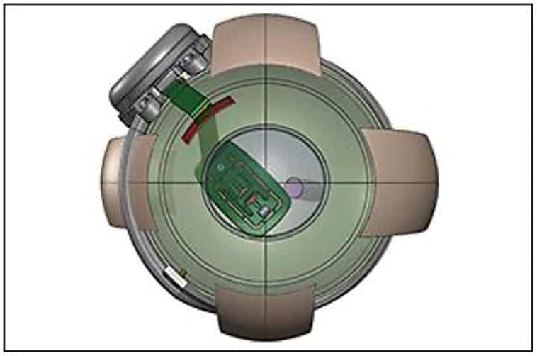
Illustration – Schematic of Argus II implantation showing the array cable entering the eye through a sclerotomy located in the superotemporal quadrant and the placement of the array over the macula. Credits: Retinal Physican
The work of Lazzi and his colleagues at USC is focused on models of nerve cells that transmit visual signals from the eye to the brain. But how does this work? We need to look into how vision occurs in the human eye in order to help us understand the computer models that underlie the research. The retina is found at the back of the eye. Light entering a healthy eye allows the lens to focus on the retina and converts into electric impulses by photoreceptors. Other cells in the retina then process these signals and pass them on to ganglion cells, which connect the retina to the brain with its long tails called axons. Axons bundle together to make up the optic nerve that delivers information to the brain.
But in a degenerative eye photoreceptors and processing die off, while retinal ganglion cells usually remain functional for longer. The Argus II targets signals directly to those specific cells. As electrical engineers, Lazzi and his colleagues’ research aims at producing a good set of electrical inputs to ganglion cells, recreating what the photoreceptors and processing cells do.
Patients with the bionic eye receive a small implant with a range of electrodes which activate when they receive a signal from a pair of special glasses that have a camera on them. Depending on the light pattern received, the camera determines the retinal ganglion cells to activate. The signals sent to the brain form a vision of black and white image made up of 60 dots.
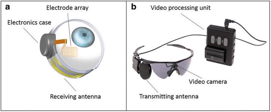
Illustration – The Argus II System. (a) Implanted components of the system. (b) External components of the system. Credits: Semantic Scholar
The device sometimes activates neighboring axons of cells of its target under certain conditions, which results in the vision of some elongated shapes instead of dots. Lazzi and his colleagues’ research addressed this issue by designing an electrical stimulation waveform that more precisely targets the cell. Modeling two subtypes of retinal ganglion cells and applying the models at the single-cell level and at the network level, the researchers found a pattern of short pulses that more precisely targets cells of interest, with less activation of neighboring axons.
Interestingly, changes in the frequency of the electrical signal result in the perception of different colors. In another study published in the journal Scientific Reports, the same modeling system was applied on the same two cell types with the goal of encoding color in the vision. Lazzi and his colleagues used the same approach and successfully developed a strategy that adjusts the signal’s frequency for the perception of the color blue.
Combining the existing models with the use of artificial intelligence can lead to advancements beyond adding colors to the bionic eye. A useful next step is to make important features such as faces or doorways stand out in a person’s vision.
“We can gift these prosthetics with intelligence, and with knowledge comes power”, Lazzi said. Although, there is still much to be done, the researchers are making great progress in the right direction with exciting advancements to follow.
References
Keck School of Medicine of USC, 2021, Advanced Computer Model Enables Improvements to “Bionic Eye” Technology, SciTechDaily, 27th April 2021, https://scitechdaily.com/advanced-computer-model-enables-improvements-to-bionic-eye-technology/
